Oral Pathology and Oral Microbiology
& Department of Dental Anatomy, Embryology & Oral Histology
& Department of Dental Anatomy, Embryology & Oral Histology
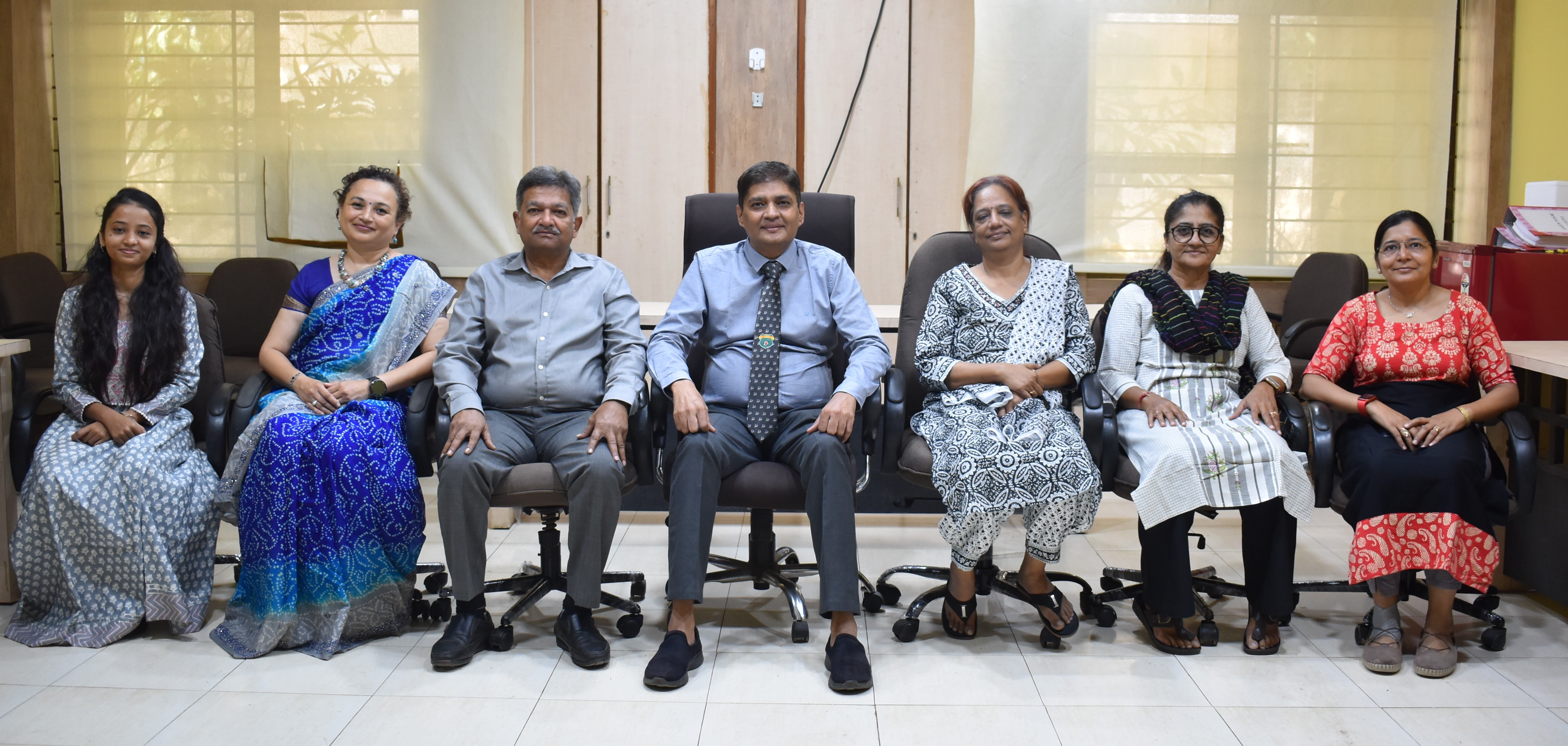
Headed by Prof. Dr. Jitendra K. Rajani, the Department strives to provide basic
knowledge of normal dental anatomy, histology and pathology for improved understanding of the disease by
increasing the diagnostic skills. Dr. Rajani has a number of national and international publications to his
name, and has earned great laurels with his successful dental practice for more than two decades now.

|
Facilities
|
- Clinic for diagnostic procedures
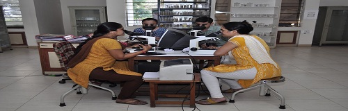
- Computer room having microscope and camera attachment along with image analysis software
- Separate sections for undergraduate and postgraduate students
- Oral pathology lab
- Partnership with Gujarat Cancer & Research Institute for the placement of postgraduate students
- Histopathology procedures lab, grossing room, processing room and seminar room with projector facility
- Separate dental anatomy and dental histology laboratory
- Well-maintained museum which has a large collection of hard and soft tissue specimens of some common as well as rare disease along with their clinical, radiographic details for students and visitors to help understand the subject at a glance
|
Client Services
|
Histopathological Examination
The Department deals with the diagnosis of variety of disorders that manifest in
the oral cavity and structures adjacent to it. Diseases with common presentation but different behaviour
need to be evaluated with microscopic examination. The surgically removed representative areas of disease in
the patient are processed, followed by the microscopic examination. Accurate diagnosis is made, based on
which treatment modalities and outcome of the procedures can be decided.
Cytology Examination
Patients having habit of tobacco chewing or smoking and ones who are more likely
to develop cancer are examined and treated by simple outpatient department procedure of cytology. The
procedure neither requires any special instrument nor is it technique sensitive. The early diagnosis of such
diseases by this method can help in early detection of cancer.
Recent Advances
The Microscope with camera attachment:
It is a helpful tool in simultaneous evaluation of microscopic image. It can be helpful for study purpose
and documentation too.
Image analysis software: It is latest image analysis software, used by scientists
for analysing the images in a simple manner. Live images can be seen and captured with additional advantage
of analysing it at the same time.
Armamentarium
- Automatic Tissue Processor
It is a series of tissue containers which contains several reagents and the embedding medium in the end. It is used for the purpose of processing of tissue sample. - Phase Contrast Microscopy
This microscopy technique is also very essential for studying structures in detail. It converts phase shifts in light and help visualises the object of interest in a better way.
- Automatic Knife Sharpener
It is an automatic tool which sharpens the knife used with the microtome. It is safe and effective method with which one can have a perfect knife edge. - Dark Field Microscopy
It is a microscopy technique in which different illumination technique is used to enhance the contrast in unstained samples.
- Manual and Semi-Automatic Microtome
It is used for accurate cutting of tissue samples at a precise thickness in a controlled and comfortable manner. - Polarising
Microscopy
A valuable diagnostic tool that helps in better understanding of the isotropic and anisotropic structures. - Stereo Microscope
Used to view enlarged images of objects without being processed.

| Prosthodontics
and Crown & Bridgework |

|
| Periodontics and Oral Implantology |
|
| Conservative Dentistry & Endodontics |
|
| Paediatric & Preventive Dentistry |
|
| Oral Medicine & Radiology |
|
| Orthodontics and Dentofacial Orthopaedics |
|
| Public Health Dentistry |
|
| Oral Pathology and Oral Microbiology & Department of Dental Anatomy, Embryology & Oral Histology |
|
| Oral & Maxillofacial Surgery |
|